May 14, 2025
Seeing More with Less: Super-Resolution Lung MRI at 0.55T
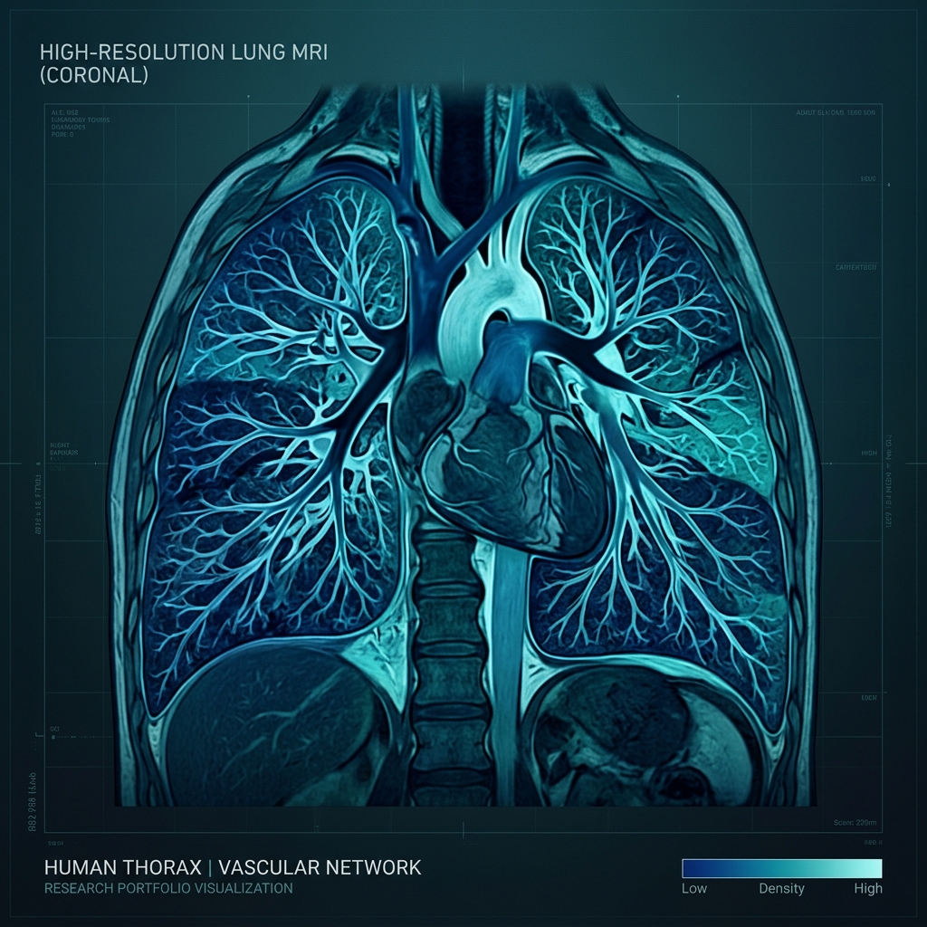
Low-field MRI is having a renaissance. Scanners operating at 0.55T are cheaper to build, cheaper to run, and — counterintuitively — often better for lung imaging: susceptibility gradients at air-tissue interfaces scale with field strength, so at low field the lung parenchyma stays visible for longer. The catch? Lower field means lower signal, which traditionally means lower resolution.
bSTAR: Free-Breathing 3D Acquisition
Our group images the lung with bSTAR (balanced steady-state free precession with stack-of-stars sampling), a sequence that lets patients breathe freely during the scan. The acquired 3D morphological images are robust, but their native resolution leaves fine structures — small airways, vessels, early fibrotic change — at the edge of visibility.
Learning to Sharpen Physics
This is where super-resolution deep learning comes in. Instead of asking the scanner for more signal, we train a network to recover high-frequency detail from the low-resolution acquisition, using paired data and physics-consistent degradation models. The key is honesty: the network must enhance what is truly there, not hallucinate plausible-looking anatomy. We constrain training with realistic point-spread functions and validate against higher-resolution references.
I presented this work as a PowerPitch at ISMRM 2025 in Hawaii. The combination of low-field hardware and learned reconstruction points to a future where high-quality lung MRI is accessible far beyond major research hospitals.