May 10, 2025
The Battle of the Breath: Overcoming Inhomogeneity in Lung MRI
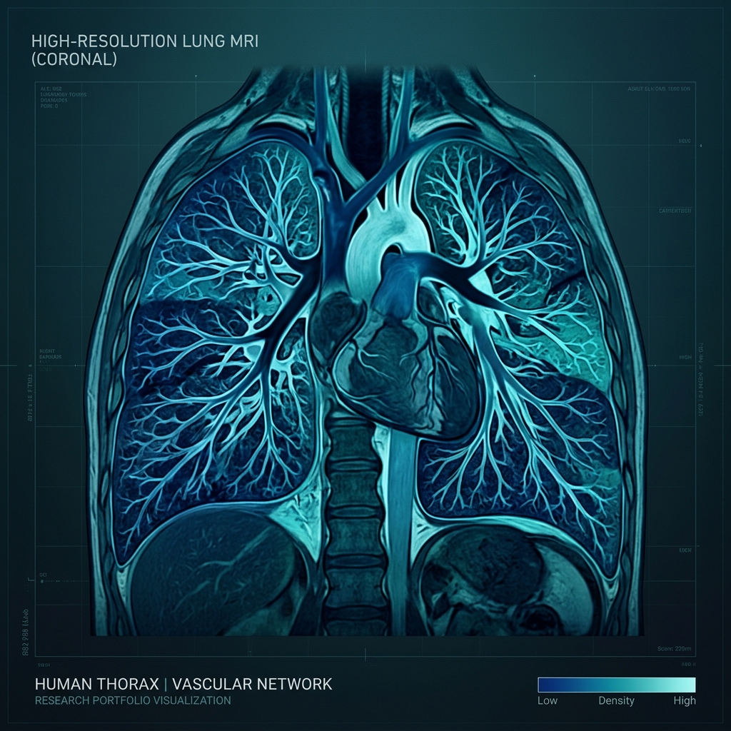
Imaging the human lung using Magnetic Resonance Imaging (MRI) is famously difficult. The lung consists of millions of microscopic air-tissue boundaries (alveoli). This interface causes rapid changes in magnetic susceptibility, leading to a highly inhomogeneous local magnetic field (\(B_0\)).
Susceptibility & Banding Artifacts
When using balanced Steady-State Free Precession (bSSFP) sequences (which offer high signal-to-noise ratios and fast scans), these field inhomogeneities cause off-resonance phase wrapping. Visually, this manifests as dark stripes—known as banding artifacts—directly across the lung parenchyma, rendering the scan clinically useless.
Active 2D Inline Shimming
To combat this, my research focuses on Active Shimming. Shimming involves adjusting auxiliary electromagnetic coils to create a counter-field that cancels out inhomogeneities. Our group developed an inline, automated 2D shimming protocol at 3T. By calculating localized field maps during a single free-breathing acquisition, we dynamically adjust the shimming gradients, mitigating banding artifacts and restoring clear morphological depictions of the lung tissue.
This advances functional lung imaging, paving the way for non-invasive tracking of chronic lung conditions.